
Article Plan: Sternal Precautions Exercises PDF
This document details a revised approach to post-sternotomy recovery, focusing on early, safe exercise protocols and challenging outdated restrictions.
Historically, sternal precautions following median sternotomy were rigidly enforced, stemming from concerns about post-operative wound instability and potential complications. These guidelines traditionally aimed to protect the sternal closure during the critical healing phase. However, contemporary research increasingly questions the necessity of such restrictive protocols.
The SAFE-ARMS study, alongside other literature reviews, suggests that early, progressive exercise – even incorporating resistance training – doesn’t demonstrably increase the risk of sternal complications. This shift in understanding necessitates a re-evaluation of standard practice, moving towards a more active and individualized rehabilitation approach. This document outlines this evolution, providing a framework for safe and effective exercise programs post-sternotomy.
Understanding Median Sternotomy
Median sternotomy is a common surgical approach for various cardiac procedures, involving a vertical incision through the sternum – the breastbone. This allows surgeons access to the heart and surrounding structures. Understanding the procedure and subsequent healing process is crucial for developing appropriate rehabilitation protocols.
The surgical procedure itself creates a temporary disruption of the chest wall’s structural integrity. Post-operatively, the body initiates a complex healing cascade, involving bone and soft tissue repair. Traditional precautions were based on assumptions about the fragility of this repair, but current evidence suggests the sternal closure possesses significant strength.
The Surgical Procedure
The median sternotomy begins with a midline incision extending from just below the neck to below the xiphoid process. The sternum is then carefully divided using a saw, providing access to the heart. Following the cardiac procedure, the sternum is re-approximated and secured with stainless steel wires.
This closure, while strong, represents a surgically created fracture. Early assumptions focused on the potential for instability, leading to restrictive post-operative protocols. However, research indicates the repair’s strength exceeds what historical precautions implied, prompting a re-evaluation of rehabilitation strategies.
Post-Operative Healing Process
Following median sternotomy, the healing process involves initial inflammation, followed by callus formation and eventual bone remodeling. This typically takes several weeks to months, but individual timelines vary. Early concerns centered on the potential for delayed healing or sternal dehiscence – a separation of the sternum – leading to significant morbidity.
Traditional protocols aimed to protect the sternum during this vulnerable period. However, emerging evidence suggests that prolonged restriction can hinder recovery, impacting functional capacity and quality of life. A more active approach, guided by careful screening and individualized exercise, is now gaining traction.
Traditional Sternal Precautions: A Historical Overview
Historically, sternal precautions stemmed from early cadaver studies and a cautious approach to post-operative care after median sternotomy. These guidelines, developed before robust clinical evidence, focused on minimizing stress on the sternal closure. Restrictions included limitations on lifting – typically 5-10 lbs – and arm movements, aiming to prevent displacement or disruption of the healing bone.
This restrictive model prioritized protection over early mobilization, believing it essential to prevent complications. However, these precautions were often described as vague and overly restrictive, hindering patients’ ability to regain functional independence promptly.
Common Restrictions Following Sternotomy
Traditionally, patients undergoing median sternotomy faced significant limitations post-operatively. Lifting restrictions were a cornerstone, commonly capped at 5-10 lbs, intended to minimize strain on the sternal wound. Arm movements were also heavily restricted, often limited in range of motion and elevation to prevent excessive force on the sternum.

These precautions aimed to protect the healing sternum, but often led to deconditioning and delayed return to activities of daily living; The vagueness of these guidelines sometimes created confusion, impacting rehabilitation program design and patient progress.
Lifting Limitations (Historically 5-10 lbs)
The 5-10 lb lifting restriction following median sternotomy has been a longstanding standard precaution. This limitation stemmed from concerns about disrupting the sternal closure and causing complications. However, recent research challenges this conservative approach, suggesting the sternal repair possesses significantly greater strength than previously assumed.
Studies indicate that the actual strength of the repair far exceeds what this weight limit implies. Consequently, many clinicians now question the necessity of such a restrictive guideline, recognizing its potential to hinder functional recovery and quality of life.

Arm Movement Restrictions
Historically, patients undergoing median sternotomy faced significant restrictions in arm movements. These limitations aimed to minimize stress on the sternum and prevent displacement of the surgical repair. Specific restrictions often included avoiding reaching, lifting the arms above shoulder height, and vigorous movements.
However, emerging evidence, like the SAFE-ARMS study, demonstrates the safety of early, controlled upper limb resistance exercises. Researchers found no clinical signs of sternal complications during these exercises, supporting a shift towards a more active rehabilitation approach. This challenges the need for prolonged, restrictive arm movement limitations.
The Rationale Behind Traditional Precautions
Traditional sternal precautions stemmed from early cadaver studies and a perceived need to protect the sternal repair. The primary concern was preventing sternal separation or displacement, which could lead to significant complications. Consequently, limitations were placed on lifting, arm movements, and overall physical activity.
These precautions were based on the assumption that the sternum was inherently unstable post-surgery and required a prolonged period of immobilization. However, recent research indicates the repair strength is considerably greater than previously believed, questioning the necessity of such restrictive protocols. The historical approach prioritized minimizing risk, even at the expense of functional recovery.
Challenging the Traditional Approach: Emerging Evidence
Growing evidence challenges the long-held beliefs underpinning traditional sternal precautions. Studies, like the SAFE-ARMS trial, demonstrate the safety and feasibility of early resistance training post-median sternotomy. Researchers observed no clinical signs of sternal complications – increased pain, clicking, or separation – during upper limb exercises.
This research aligns with prior findings supporting active, unweighted movements in cardiac surgery patients. The emerging consensus favors an active participatory model of cardiac rehabilitation, prioritizing early engagement in exercise to reduce pain and improve daily function. This shift moves away from restrictive protocols based on outdated cadaver studies.
The SAFE-ARMS Study Findings
The SAFE-ARMS study rigorously investigated the safety of early resistance training following median sternotomy. Researchers found no clinical indications of sternal complications – such as increased pain, crepitus, or palpable sternal separation – during the performance of six upper limb resistance exercises. These findings strongly suggest that early, controlled resistance training is well-tolerated by patients.
The study supports a departure from historically restrictive protocols. It reinforces the idea that active engagement in exercise, rather than prolonged restriction, can facilitate recovery and improve functional capacity. This evidence contributes to a growing body of literature advocating for an active participatory model of cardiac rehabilitation.
Safety of Early Resistance Training
The SAFE-ARMS study demonstrated a notable absence of sternal complications when implementing early resistance training post-median sternotomy. Specifically, researchers observed no significant increases in pain beyond baseline levels during exercise. Furthermore, there were no reports of concerning signs like clicking, crepitus, or increased sternal motion.
This finding aligns with previous research supporting the safety of active, unweighted upper limb and trunk movements in cardiac surgery patients. The study provides compelling evidence that early, controlled resistance exercise doesn’t compromise sternal stability, paving the way for more active rehabilitation programs.
Absence of Sternal Complications in the Study
Crucially, the SAFE-ARMS study reported zero clinical indications of sternal instability or failure amongst participants engaged in early resistance exercises. Researchers meticulously monitored for signs such as heightened pain levels, audible clicking or crepitus, and palpable sternal separation – none were detected.
This consistent lack of adverse events reinforces the growing body of evidence questioning traditionally restrictive postoperative protocols. The study’s findings suggest that a more active, participatory approach to cardiac rehabilitation is not only feasible but also safe, potentially accelerating recovery and improving patient outcomes.

Sternal Strength and Repair Integrity
Research indicates the sternal repair post-median sternotomy possesses considerably greater strength than previously assumed by historical precautions. Studies challenge the long-held belief that even minimal weight lifting – like the 5-10 lb restriction – is necessary to protect the healing sternum.

Evidence suggests the biomechanical integrity of the sternal closure is robust enough to withstand significantly more stress than traditionally acknowledged. This understanding supports a shift towards individualized rehabilitation programs, tailored to patient-specific factors, rather than universally applied, overly cautious limitations.

Factors Influencing Sternal Healing
Sternal healing isn’t uniform; it’s significantly impacted by both patient-specific characteristics and surgical technique. Patient-related risks include pre-existing conditions like osteoporosis, diabetes, and obesity, alongside smoking status and nutritional deficiencies. Surgical considerations encompass the chosen closure technique – wiring versus newer alternatives – and the surgeon’s experience.
Brocki et al.’s literature review highlights these contributing factors to sternal complications. Individualized assessment of these elements is crucial for determining appropriate activity levels and exercise progression post-sternotomy, moving away from generalized precautions.
Patient-Specific Risk Factors
Numerous patient characteristics can impede optimal sternal healing. Conditions like osteoporosis significantly weaken bone density, increasing fracture risk. Diabetes impairs wound healing and immune function, while obesity adds mechanical stress to the sternum. Smoking compromises blood flow and tissue oxygenation, hindering repair.
Nutritional deficiencies, particularly Vitamin D and protein, also play a role. Pre-existing lung disease can increase post-operative pain and limit exercise tolerance. Thorough pre-operative assessment of these factors is vital for tailoring rehabilitation programs and mitigating potential complications.
Surgical Technique Considerations
The method of sternal closure profoundly impacts healing. Utilizing robust suture techniques, like figure-of-eight configurations, enhances stability. Minimizing sternal retraction time during surgery reduces tissue trauma. Employing sternal retractors with appropriate padding prevents excessive pressure.
Careful hemostasis (control of bleeding) is crucial to avoid hematoma formation, which can impede healing. The surgeon’s experience and adherence to best practices are paramount. Considering alternative closure methods, such as sternal banding, may be appropriate in high-risk cases, optimizing structural integrity.
Early Screening for Sternal Complications
Proactive assessment is vital for identifying patients at risk. Palpation of the sternum detects instability or abnormal motion. Monitoring pain levels, specifically pain exacerbated by movement, is crucial. Visual inspection reveals signs of wound dehiscence or hematoma.
Assessing for clicking or crepitus during arm movements indicates potential issues. Early screening allows for tailored exercise programs. Prompt identification enables timely intervention, preventing progression to more serious complications. A comprehensive evaluation guides safe and effective rehabilitation, optimizing patient outcomes.
Individualized Exercise Programs
Moving beyond blanket restrictions, programs should be tailored to each patient’s healing trajectory. Initial assessments determine baseline strength and stability. Progression is guided by pain levels and clinical findings, not arbitrary timelines. Early phases focus on gentle range of motion and diaphragmatic breathing.
Resistance training is introduced cautiously, starting with light weights and high repetitions. Functional activities, like reaching and lifting, are gradually incorporated. Regular monitoring ensures safety and effectiveness. Individualized plans maximize recovery and return to daily living, promoting an active participatory model.
Recommended Exercises – Phase 1 (Early Post-Op)
Phase 1 prioritizes gentle movements to restore basic function and minimize discomfort. Ankle pumps and calf stretches promote circulation. Deep breathing exercises enhance lung capacity and reduce the risk of pulmonary complications. Shoulder shrugs and pendulum exercises initiate upper limb mobility without stressing the sternum.
Gentle elbow flexion and extension maintain range of motion. Wrist rotations improve circulation in the hands. Focus is on pain-free movement, avoiding any activities that cause sternal clicking, crepitus, or increased pain. Progression is slow and deliberate, guided by patient tolerance.
Recommended Exercises – Phase 2 (Progressive Strengthening)
Phase 2 builds upon Phase 1, introducing light resistance to enhance strength and endurance. Bicep curls and triceps extensions with resistance bands begin upper limb strengthening. Shoulder external rotations and rows further improve shoulder function. Wall push-ups offer a controlled chest exercise.
Trunk stabilization exercises, like seated twists and pelvic tilts, improve core strength. Emphasis remains on proper form and avoiding sternal stress. Resistance is gradually increased as tolerated, monitoring for any signs of discomfort or instability. Progression is individualized based on the SAFE-ARMS study findings.
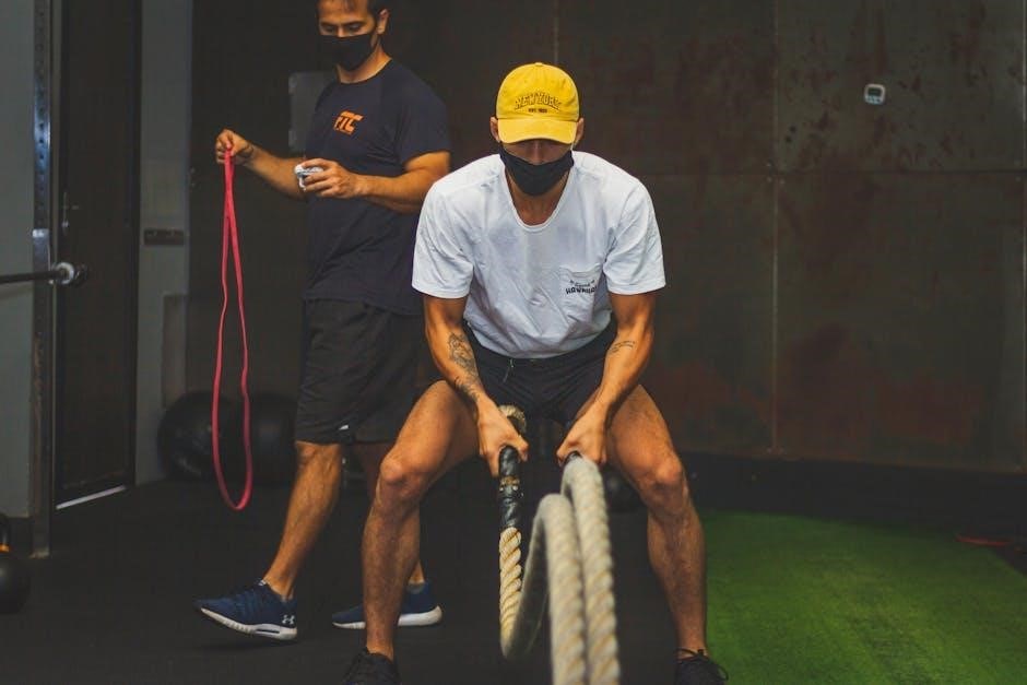
Upper Limb Resistance Exercises
These exercises are crucial for regaining upper body function post-sternotomy, guided by the SAFE-ARMS study’s safety data. Begin with light resistance bands for bicep curls, triceps extensions, and shoulder presses. Focus on controlled movements, avoiding forceful exertions or reaching. Progress gradually, increasing resistance as tolerated, while closely monitoring for any sternal discomfort.
Exercises like rows and external rotations strengthen supporting muscles. Prioritize proper form over weight, ensuring no clicking, crepitus, or increased pain. Individualized progression is key, respecting patient-specific healing rates and risk factors.
Trunk Stabilization Exercises
Strengthening core muscles is vital for overall stability and reducing strain on the sternum. Begin with gentle abdominal bracing and pelvic tilts, focusing on engaging deep core muscles without excessive movement. Progress to seated twists and side bends, maintaining a neutral spine and controlled pace. Avoid exercises that heavily load the trunk or involve forceful twisting initially.
Exercises like bird-dogs and dead bugs promote core stability. Monitor for any sternal discomfort or increased pain during these movements. Individualized progression is essential, adapting to the patient’s tolerance and healing progress.
Activity Recommendations Based on Literature Review
Current literature suggests a shift away from overly restrictive precautions. Brocki et al.’s review highlights factors influencing sternal complications, informing tailored activity guidelines. Early, controlled activity is now favored over prolonged rest. Lifting limitations should be individualized, considering repair strength—often exceeding the traditional 5-10 lb restriction.

Focus on functional movements mimicking daily tasks. Gradual progression is key, monitoring for pain or instability. Active participation in cardiac rehabilitation, with resisted exercises, is encouraged to optimize recovery and return to function.
Moving Towards an Active Participatory Model of Cardiac Rehabilitation
The SAFE-ARMS study strongly supports an active participatory model, demonstrating the safety of early resistance training post-sternotomy. This approach prioritizes patient engagement and functional recovery over historical restrictions. Traditional protocols, derived from cadaver studies, are increasingly questioned. Early active exercise reduces pain, facilitates daily living activities, and optimizes overall recovery.
Cardiac rehabilitation should focus on individualized programs, empowering patients to actively participate in their healing process, rather than adhering to blanket limitations.
Benefits of Early Active Exercise
Early active exercise following median sternotomy offers significant advantages, moving beyond restrictive postoperative protocols. The SAFE-ARMS study revealed no increase in sternal complications with early resistance training, bolstering confidence in this approach. Active participation reduces pain and improves functional capacity, enabling patients to return to activities of daily living more quickly.
This proactive model optimizes recovery, contrasting with historically vague and overly restrictive precautions. Individualized programs enhance patient well-being and promote a faster, more complete return to a normal lifestyle.
Potential Risks and Monitoring
While early active exercise is beneficial, careful monitoring is crucial. Potential risks include discomfort or pain, though the SAFE-ARMS study showed no clinical signs of sternal complications with the tested exercises; Regular assessment for increased pain, clicking, crepitus, or sternal separation is essential.
Healthcare professionals should screen for patient-specific risk factors and adjust exercise programs accordingly. Close observation during exercise sessions allows for prompt intervention if needed, ensuring patient safety and optimizing recovery outcomes.
Traditional sternal precautions, rooted in early cadaver studies, appear overly restrictive given current evidence. The SAFE-ARMS study and literature reviews demonstrate the safety and feasibility of early resistance training, challenging the historical 5-10 lb lifting limit.
An active participatory model of cardiac rehabilitation, prioritizing individualized exercise programs and early engagement, is recommended. This approach facilitates faster recovery, improves activities of daily living, and optimizes patient outcomes. Continued research and monitoring are vital to refine these revised guidelines.